#PDD#
Protein
Drug
Discovery
“百聞は一見に如かず”
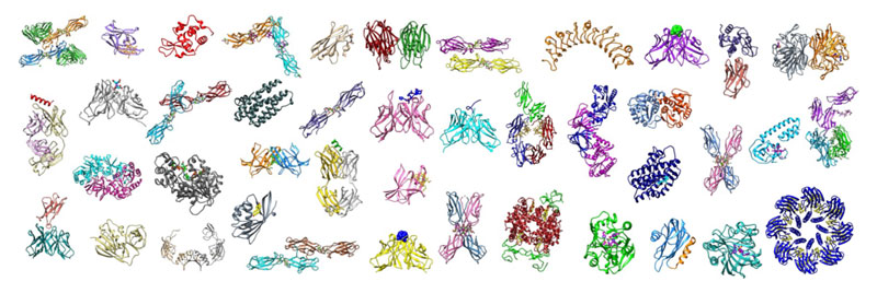
当研究室では、NMR、X線結晶構造解析、クライオ電子顕微鏡などの高分解能技術を活用して標的蛋白質の構造を決定する(=見る)研究を推進しています。私たちがこれまで構造決定してきた蛋白質は多岐にわたり、多様な分子量・蛋白質機能・薬剤としての可能性を有するものです。このような生体高分子の構造を決定することによってその形状・立体配置を可視化できるのは勿論、薬剤やリガンド・基質の蛋白質に対する結合様式を解明することも可能になります。それによって例えば、蛋白質表面にある薬剤結合ポケットを解析し、部位特異的変異導入と併用することで化合物スクリーニングから得られたヒット化合物を最適化する、いわゆるQSAR(Quantitative Structure-Activity Relationship)と呼ばれる研究を強力に推進できます。さらには、抗原に結合する抗体の構造を解明できればその相互作用を理解し、よりよいワクチンのデザインへ研究が展開していくことも期待されます。このように、構造解析は当研究室における研究活動の中心軸となります。当研究室に参画する暁には、構造取得に関係する実験手技を体得し、私たちの構造ギャラリーに新たな蛋白質構造を加えていくことになることでしょう。
"Seeing is believing" .
In our laboratory we employ high-resolution techniques such as Nuclear Magnetic Resonance (NMR), X-ray crystallography, and Cryo-EM, to determine (“to see”) the structures of proteins of interest. Our protein gallery includes proteins of different sizes, biological functions, and therapeutic potential. Determining the structure of a macromolecule not only provides a clear picture of its shape and three-dimensional arrangement, but it is also the preferred method to observe the binding of drugs (or ligands, substrates, etc...) to proteins. Analyzing the pockets on the surface of the protein in combination with site-directed mutagenesis helps to design better hit-candidates after the library screening step, and also support the subsequent QSAR stages. In addition, revealing the structures of antibodies bound to their antigens is a very powerful method to understand their interaction, and this method may be employed in vaccine design. For all these reasons, structural analysis becomes one of the main pillar of our research. Our students are expected to gain a sophisticated understanding of these techniques and their analytical tools.